Special Issue: Echinoderms - from the early past to the near future. A tribute to Hans Hess on his 80th birthday
- Published:
Pentasteria? splendida, a new Early Cretaceous astropectinid starfish from northern Germany
Swiss Journal of Palaeontology volume 130, pages 123–127 (2011)
Abstract
A new species of early Hauterivian (Early Cretaceous) astropectinid starfish, Pentasteria? splendida, is recorded from strata assigned to the Endemoceras amblygonium ammonite Zone at Engelbostel, near Hannover (northern Germany). Although based solely on a lightly pyritised proximal arm fragment (most likely the result of a predator attack, which is also suggested by possible bite marks), preservation is otherwise excellent, with numerous spines on inferomarginals and adambulacrals retained. Distinguishing features include a distal row of three equal-sized spines on the oral surface of inferomarginals, plus two oblique rows of horseshoe-shaped spine bases on their lateral surface bearing spines of variable length, as well as the absence of large spine pits on superomarginals and adambulacrals. Two oral intermediate ossicles close to the disc margin apparently possess larger spine bases. For the time being, assignment to the family Astropectinidae is based on the occurrence of multiple spines on inferomarginal ossicles and paxilliform adoral ossicles, but placement in the genus Pentasteria is tentative, because superomarginal ossicles have closely spaced small granules rather than scale-like spinelets.
Introduction
As recently shown by Villier (2010), the record of Early Cretaceous asteroid species from Europe and North Africa is comparatively poor, albeit severely biased. On the basis of dissociated ossicles from the Barremian (Early Cretaceous) of Drôme (southeast France), Villier (2010) described astropectinids, stauranderasterids, goniasterids, ?sphaerasterids, a valvatidan clade of uncertain affinities and ?forcipulatids, inclusive of two new species. With these additions, around 20 species are now known (compare Villier (2010), Table 1). Taxa that are more or less coeval with the present record from northern Germany include the following:
-
*Comptoniaster godeti Breton 1992, a goniasterid from the Hauterivian of Isère, France;
-
*Pycinaster sp., another goniasterid from the same level and locality;
-
*Chaetasterina gracilis Hess 1970, a chaetasterid from the Hauterivian of Sainte-Blaise, Switzerland;
-
*Protothyraster priscus (de Loriol 1874), an ophidiasterid from the same level and locality;
-
*Coulonia neocomiensis de Loriol 1874 (= Cuneaster hauteriviensis Hess 1955), an astropectinid from the Hauterivian of Neuchâtel, Switzerland and Drôme, France;
-
*Coulonia platyspina Hess and Blake 1995, an astropectinid from the Barremian of Agadir, Morocco;
-
*Pentasteria sp., an astropectinid from the Hauterivian of Sainte-Blaise, Switzerland (see Hess 1970) and;
-
*indeterminate benthopectinids from the Hauterivian of Isère, France.
To this list should be added some preliminary records of well-preserved early Valanginian asteroids from the Platylenticeras heteropleurum and P. robustum ammonite zones at Sachsenhagen, northern Germany, mostly contained in private collections [see Pockrandt and Meyer (1986); Fischer (1992); Zawischa (1992)]. Unfortunately, these have not yet been properly described and named.
As far as we are aware, the first record of an early Hauterivian asteroid from Engelbostel is a partial specimen, retaining portions of at least two arms, in private hands (B. Dose Collection). It was briefly discussed and illustrated by Weitschat (1984, pp. 95, 96, unnumbered figure), who compared it with representatives of the genera Cottreauaster C.W. Wright 1951 and Comptonia Gray 1840. Although we have been unable to trace this specimen ourselves, the schematic drawing in Weitschat (1984) does show a certain resemblance to the present record (i.e. nature of the marginal ossicles, width and length of arms), and may actually refer to the same astropectinid which is here described as a new species.
Systematic palaeontology
-
Order Paxillosida Perrier 1884
-
Family Astropectinidae Gray 1840
-
?Genus Pentasteria Valette 1929
-
Type species: Pentasteria boisteli Valette 1929, by monotypy.
-
Pentasteria? splendida n. sp. (Fig. 1)
Fig. 1 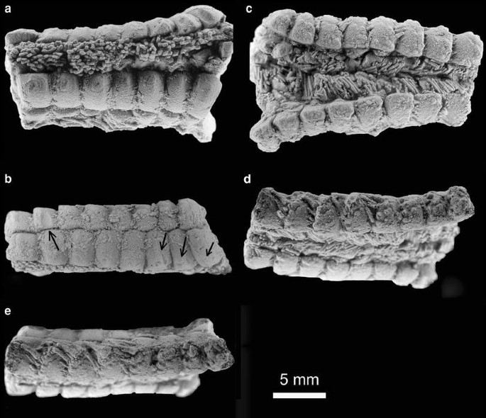
Pentasteria? splendida n. sp., GPIMH 3100 (holotype), Endemoceras amblygonium ammonite Zone (early Hauterivian, Early Cretaceous) of Engelbostel (Hannover area, northern Germany), in various aspects. a, aboral view; b, lateral view (note possible bite marks; arrowed); c, oral view; d. oblique oral/lateral view to show horseshoe-shaped spine bases; e, lateral/oral view. Specimen whitened with ammonium chloride prior to photography
-
*Pentasteria sp. Neumann(2010), p. 35, Figs. 1–3.

Material
GPIMH 3100 (leg./don. Norbert Nordmeyer), the holotype and sole specimen known to date, in the collections of the Geologisch-Paläontologisches Institut und Museum der Universität Hamburg.
Locality and stratigraphy
Former clay pit Engelbostel, about 5 km north of Hannover, northern Germany; earliest Hauterivian (lower Endemoceras Beds; Endemoceras amblygonium ammonite Zone; see Mutterlose (1998), pp. 73, 74, Fig. 34; Mutterlose et al. (1998, Fig. 4).
Derivation of name
Named after Norbert Nordmeyer, the name ‘Norbert’ in Germanic language meaning ‘the splendid one from the north’.
Diagnosis
Medium-sized astropectinid, with narrow, long arms; superomarginals with close-set granule pits only, lacking large spine pits; inferomarginals with distal row of three equal-sized spines, plus two oblique lateral rows of larger horseshoe-shaped spine bases to which are attached spines of variable length; adambulacrals lacking large spine pits, oral intermediate ossicles close to disc margin apparently with large spine base.
Description
Fragmentary proximal arm (eight supero- and eight inferomarginals); greatest length (as preserved) 18.2 mm, distal width 7.8 mm, proximal width 10.8 mm; pyritised and compressed laterally, making measurements of arm width approximate; comparison with well-preserved astropectinids such as a specimen of Pentasteria longispina Hess 1968 (see Hess 1975, p. 1), suggests a medium-sized individual, with a major radius (R) of c. 80 mm and a minor one (r) of c. 20 mm. Superomarginals near disc margin (Fig. 1a, right-hand side) rather tall (ossicular angle c. 48 degrees), progressively becoming longer more distally and more quadrangular in outline; length between 2.0 and 2.2 mm proximally to 2.4–2.5 (and more) distally (Fig. 1a, b); only with closely spaced, small granule pits; in places, diminutive, roundish and flat granules preserved (Fig. 1a, b) where ossicles abut; aboral surface of superomarginals bulging, ridge-like but merging smoothly into remainder of ossicle (Fig. 1a, b), flatter more distally and corrugated to some extent (Fig. 1a, left-hand side), but lacking large spine pits. In lateral view (Fig. 1b), supero- and inferomarginals alternate in the proximal and median arm portion, but are opposed more distally; proximal facets of infero- and superomarginals with rather shallow intermarginal fascioles. Proximal inferomarginals more cuneate (Fig. 1c), becoming more quadrangular distally (Fig. 1c) and there with at least three equal-sized, close-set, striate spines (length c. 50% of marginal length) on oral surface, near distal margin (Fig. 1c) and up to six (in two oblique rows) on lateral surface, three stouter, the others of variable length, all finely striate; the longest spine at least corresponds to the length of an inferomarginal ossicle; spine bases horseshoe shaped, one row of four, near-equal sized spine bases distal margin oblique towards proximal–intermarginal corner, second row of two slightly smaller, horseshoe-shaped spine bases distal to first row (Fig. 1c–e). Ambulacrals not visible; aboral ossicles quadrangular, blocky (Fig. 1a), near-flat to slightly bulbous, with smooth surface; paxilliform adoral ossicles close set, at least 9–11 rows abreast, pillar like, c. 0.5 mm in length, with only slightly widened and rounded base (Fig. 1a). Adambulacrals blocky, with uneven outer surface, smooth, alternating in opposite rows, with close cover of similar-sized spines, straight to lightly curved (Fig. 1c, d), striate and up to 2 mm in length, and up to 5–6 per ossicle. No larger spines, but a row of oral intermediate ossicles proximally with 3–4 smaller spines, and even more proximal ones, at disc margin (Fig. 1c, lower right-hand side), apparently with a single, large spine base.
On some marginal ossicles (Fig. 1b, arrowed) tooth marks are visible [compare Neumann (2000); Zatoń et al. (2007)], which would indicate a predator attack and might also explain the fragmentary preservation of this individual, which otherwise retains many skeletal appendages.
Discussion
As listed by Villier (2010, Table 1), there is only a single previous record of a species of Pentasteria from Hauterivian strata. Hess (1970, p. 1082, pl. 3, Fig. 3; p. 4, Fig. 3) described P. (Archastropecten) sp. from the early Hauterivian of Sainte-Blaise near Neuchâtel (Switzerland), on the basis of what appeared to be a juvenile specimen (R = 8 mm; r = 4 mm). Although details of spines and marginal ornament could not be fully assessed, Hess (1970) suggested that this could be likened to common dissociated, coeval astropectinid marginals from the Waadtland (L’Auberson near Ste Croix) and the Neuenburger (Le Landeron) Jura Chain, and referred to by him as P. (A.) cf. huxleyi. However, as illustrations in Hess (Hess 1955, p. 87, Figs. 23–37) and Hess (Hess 1975, pp. 29, 34, pl. 8, Figs. 13–14) show, this form has much better-developed intermarginal fascioles, superomarginals are much lower than those of P.? splendida n. sp. and have a much coarser ornament; in addition, inferomarginals have an utterly different arrangement of spine bases.
There are, however, two additional records that need to be considered here. Firstly, from the Hauterivian of Neuchâtel, de Loriol (1874, p. 6, pl. 1, Fig. 1) described a new species, Astropecten desori, on the basis of a near-complete individual (R = 74 mm, r = 20 mm). Hess (1955, p. 32) noted that in this form the ‘ventrolateralia’ (= oral indeterminate ossicles, as here understood) were not developed far beyond the disc, meaning also that, proximally, the arms were already narrow. The marginals are described as low, and remains of inferomarginal spines suggest a length equal to the width of a plate. In these features, as well as in the coarse ornament of marginals (see de Loriol 1874, pl. 1, Fig. 1d), de Loriol’s species differs from P.? splendida n. sp. Secondly, Astropecten carthusiae Hérenger 1944 (p. 38, text-Figs. 4–9; pl. 1, Figs. 1–4), from the late Valanginian of Grande Chartreuse (France), which, according to Hess (1955, pp. 54, 55) has a single row of narrow ‘ventrolateral’ ossicles over the entire arm length and wide, low marginals and adambulacrals. Here too, confusion with the new species can be ruled out on the basis of these features.
In conclusion, the new species is provisionally assigned to the genus Pentasteria, from which it differs in having only small, close-set granules, rather than scale-like spinelets, on the superomarginals. The absence of large spine pits on those superomarginal recalls certain species of the subgenus Archastropecten Hess 1955 (sensu Hess 1960a; type species: Astropecten huxleyi T. Wright 1862), although both taxa would appear to be in need of a modern revision. As demonstrated on a number of recent occasions (see e.g. Blake 1986, 1989), some Jurassic and Early Cretaceous ‘astropectinids’ may reveal characters that are more typical of goniasterids, suggesting that those species should be transferred from the order Paxillosida to the order Valvatida. Be that as it may, in our view P.? splendida n. sp. has an inferomarginal spine canopy which is typically astropectinid in nature, although it appears more complex than in members of Pentasteria that have been described so far (see e.g. Hess 1955, 1960b, 1987) and the erection of a new genus might be warranted once better-preserved material becomes available. Unfortunately, ambulacrals are not visible; we have refrained from further preparation of this unique specimen, which adds to the meagre record of Early Cretaceous asteroids, so as not to cause irreparable damage to it.
References
Blake, D. B. (1986). Some new post-Paleozoic sea stars (Asteroidea: Echinodermata) and comments on taxon endurance. Journal of Paleontology, 60, 1103–1119.
Blake, D. B. (1989). Asteroidea: functional morphology, classification and phylogeny. In M. Jangoux, & J. M. Lawrence (Eds.), Echinoderm studies, Rotterdam/Brookfield, (3, pp. 179–223).
Breton, G. (1992). Les Goniasteridae (Asteroidea, Echinodermata) jurassiques et crétacés de France: taphonomie, systématique, paléobiogéographie, évolution. Bulletin trimestriel de la Société géologique de Normandie et des Amis du Muséum du Havre 78 (hors série), (pp. 1–590).
de. Loriol, P. (1874). Description de quelques astérides du terrain Néocomien des environs de Neuchâtel. Mémoires de la Société des Sciences naturelles de Neuchâtel, 5, 3–19.
Fischer, R. (1992). Ziegeleitongrube Sachsenhagen–ein Nachruf. Arbeitskreis Paläontologie Hannover, 20, 25–32.
Gray, J. E. (1840). A synopsis of the genera and species of the class Hypostoma (Asterias Linn.). Annals and Magazine of Natural History (1)6, pp. 175–184, 275–290.
Hérenger, L. (1944). Sur la présence d’astéries (Astropecten carthusiae nov. sp.) dans le Valanginien de la Chartreuse (Isère). Travaux du Laboratoire de Géologie de la Faculté des Sciences de Grenoble, 24, 33–47.
Hess, H. (1955). Die fossilen Astropectiniden (Asteroidea). Neue Beobachtungen und Übersicht über die bekannten Arten. Schweizerische Paläontologische Abhandlungen, 71, 1–113.
Hess, H. (1960a). Über die Abgrenzung der Astropectiniden-Gattungen Pentasteria Valette und Archastropecten Hess. Eclogae geologicae Helvetiae, 53, 329–331.
Hess, H. (1960b). Pentasteria (Archastropecten) procera n. sp. (Asteroidea, Astropectinidae) aus dem Bajocien von Cheltenham, England. Eclogae geologicae Helvetiae, 53, 321–334.
Hess, H. (1968). Ein neuer Seestern (Pentasteria longispina n. sp.) aus den Effingerschichten des Weissensteins (Kt. Solothurn). Eclogae geologicae Helvetiae, 61, 607–614.
Hess, H. (1970). Schlangensterne und Seesterne aus dem oberen Hauterivien “pierre jaune” von St. Blaise bei Neuchâtel. Eclogae geologicae Helvetiae, 63, 1069–1091.
Hess, H. (1975). Die fossilen Echinodermen des Schweizer Juras. Seesterne, Schlangensterne, Seelilien, Seeigel, Seewalzen. Veröffentlichungen aus dem Naturhistorischen Museum Basel, 8, 1–130.
Hess, H. (1987). Neue Seesternfunde aus dem Dogger des Schweizer Juras. Eclogae geologicae Helvetiae, 80, 907–918.
Hess, H., & Blake, D. B. (1995). Coulonia platyspina n. sp., a new astropectinid sea star from the Lower Cretaceous of Morocco. Eclogae geologicae Helvetiae, 88, 777–788.
Mutterlose, J. (1998). C 2.5 Hauterivian of Engelbostel. In J. Mutterlose, A. Bornemann, S. Rauer, C. Spaeth, & C. J. Wood, (Eds.), Key localities of the northwest European Cretaceous. Bochumer geologische und geotechnische Arbeiten (48, pp. 73–74).
Mutterlose, J., Wood, C. J., & Ernst, G. (1998). The Lower and upper cretaceous of the Hannover-Braunschweig area (NW-Germany). Part C. In J. Mutterlose, A. Bornemann, S. Rauer, C. Spaeth, & C. J. Wood, (Eds.), Key localities of the northwest European Cretaceous. Bochumer geologische und geotechnische Arbeiten (48, pp. 39–51).
Neumann, C. (2000). Evidence of predation on Cretaceous sea stars from northwest Germany. Lethaia, 33, 65–70.
Neumann, C. (2010). Pentasteria sp., ein Kammseestern (Astropectinidae) aus dem Unter-Hauterive von Engelbostel. Arbeitskreis Paläontologie Hannover, 38, 35–40.
Perrier, E. (1884). Mémoire sur les étoiles de mer recueillies dans la mer des Antilles et du Golf du Mexique. Nouvelles Archives du Muséum d’Histoire naturelle Paris, 6, 127–276.
Pockrandt, W., & Meyer, D. (1986). Neue Funde unserer Mitglieder. Arbeitskreis Paläontologie Hannover, 14, 86–89.
Valette, A. (1929). Note sur quelques stellerides jurassiques du Laboratoire de Géologie de la Faculté des Sciences de Lyon. Travaux du Laboratoire de Géologie de la Faculté des Sciences du Lyon, 16, 5–39.
Villier, L. (2010). Asteroids from Barremian calciturbidites of the Serre de Bleyton (Drôme, SE France). Annalen des Naturhistorischen Museums in Wien, A112, 701–732.
Weitschat, W. (1984). Ein seltener Seesternfund aus dem Hauterive von Engelbostel. Arbeitskreis Paläontologie Hannover, 12, 95–96.
Wright, C. W. (1951). Proposed use of the plenary powers to designate a lectotype for the nominal species “Asterias quinqueloba” Goldfuss, 1831 (Class Asteroidea) in harmony with currently accepted nomenclatorial practice. Bulletin of Zoological Nomenclature, 6, 106–110.
Wright, T. (1862). Monograph on the British fossil Echinodermata of the Oolitic formations, Volume II. steroidea and Ophiuroidea. AMonograph of the Palaeontographical Society London, 1(1861), 1–130.
Zatoń, M., Villier, L., & Salamon, M. A. (2007). Signs of predation in the Middle Jurassic of south-central Poland: evidence from echinoderm taphonomy. Lethaia, 40, 139–151.
Zawischa, D. (1992). Fossilien aus der Tongrube Sachsenhagen. Arbeitskreis Paläontologie Hannover, 20, 33–50.
Acknowledgments
We thank Wolfgang Weitschat (formerly Geologisch-Paläontologisches Institut und Museum der Universität Hamburg) for providing access to collections in his care and Christian Meyer (Naturhistorisches Museum Basel) and a second, anonymous reviewer for pertinent comments on an earlier draft of the typescript; one of us (JWMJ) acknowledges the financial support of the European Commission’s Research Infrastructure Action through the SYNTHESYS Project (DE-TAF-4567), which allowed him to work at the Museum für Naturkunde (Berlin) for two weeks in December 2008.
Author information
Authors and Affiliations
Corresponding author
Rights and permissions
About this article
Cite this article
Neumann, C., Jagt, J.W.M. Pentasteria? splendida, a new Early Cretaceous astropectinid starfish from northern Germany. Swiss J Palaeontol 130, 123–127 (2011). https://doi.org/10.1007/s13358-010-0011-2
Received:
Accepted:
Published:
Issue Date:
DOI: https://doi.org/10.1007/s13358-010-0011-2